
Embark on a scientific journey with our Time for Mitosis Lab Answer Key, a comprehensive guide that unravels the intricate process of cell division. Dive into the depths of mitosis, its significance, and the captivating stages that orchestrate the creation of new cells.
This meticulously crafted resource provides a clear understanding of the materials and equipment required for your lab, ensuring a seamless experimental procedure. Prepare and observe mitosis slides with precision, guided by our expert instructions. Witness the remarkable dance of chromosomes as they align, divide, and migrate, captured in detailed observations and results.
Introduction

Mitosis is a fundamental process in cell division, essential for growth, development, and tissue repair in eukaryotic organisms. It ensures the accurate distribution of genetic material to daughter cells, maintaining the chromosome number and genetic integrity within a population.
Mitosis occurs in several distinct stages, each characterized by specific chromosomal behaviors and cellular changes. These stages include prophase, prometaphase, metaphase, anaphase, and telophase, followed by cytokinesis, which divides the cytoplasm and completes cell division.
Stages of Mitosis
During prophase, the chromatin condenses into visible chromosomes, and the nuclear envelope breaks down. The spindle apparatus, consisting of microtubules, begins to form.
In prometaphase, the mitotic spindle fibers attach to the chromosomes at their kinetochores, and the chromosomes align at the metaphase plate.
In metaphase, the chromosomes are aligned at the metaphase plate, ensuring that each daughter cell receives an identical set of chromosomes.
In anaphase, the sister chromatids of each chromosome separate and move to opposite poles of the cell.
In telophase, the chromosomes reach the poles, and the nuclear envelope reforms around each set of chromosomes. Cytokinesis follows, dividing the cytoplasm and completing cell division.
Materials and Equipment
This lab requires specific materials and equipment to effectively observe and study mitosis in plant cells.
The essential materials include:
- Light microscope: A compound light microscope with a magnification range of 400x to 1000x is necessary for observing the stages of mitosis clearly.
- Microscope slides: Clean and grease-free microscope slides are required to prepare and view the plant material.
- Cover slips: Thin, square or rectangular cover slips are used to cover the plant material on the slide, creating a thin layer for observation.
- Onion root tips: Freshly cut onion root tips serve as the plant material for studying mitosis. They contain actively dividing cells, making them suitable for observing the different stages of mitosis.
- Fixative (acetic acid and ethanol): A fixative solution is used to preserve and harden the plant material, making it easier to prepare slides.
- Hydrochloric acid (HCl): Dilute HCl is used to soften the cell walls of the plant material, allowing better penetration of stains.
- Acetocarmine stain: Acetocarmine is a vital stain that selectively binds to the chromosomes, making them visible under the microscope.
- Forceps: Forceps are used to handle the onion root tips and other materials during slide preparation.
- Dissecting needle: A dissecting needle is used to gently tease apart the onion root tips, releasing individual cells for observation.
Experimental Procedure: Time For Mitosis Lab Answer Key
The experimental procedure for preparing and observing mitosis slides involves several steps. These steps include preparing the root tip squashes, staining the slides, and observing them under a microscope.
To prepare the root tip squashes, onion root tips are collected and treated with a hypotonic solution to swell the cells. The root tips are then fixed in a fixative solution to preserve their structure. The fixed root tips are then squashed onto a microscope slide using a coverslip.
This process spreads out the cells and makes them easier to observe under a microscope.
The slides are then stained with a dye that binds to DNA. This allows the chromosomes to be visualized under a microscope. The stained slides are then observed under a microscope using different magnifications to identify the different stages of mitosis.
Microscope Use, Time for mitosis lab answer key
When using a microscope to observe mitosis slides, it is important to use the correct magnification and lighting. The magnification should be high enough to clearly see the chromosomes, but not so high that the image is blurry. The lighting should be bright enough to see the chromosomes, but not so bright that it damages the cells.
Identifying Mitosis Stages
The different stages of mitosis can be identified by the appearance of the chromosomes. In prophase, the chromosomes are long and thin. In metaphase, the chromosomes are lined up in the center of the cell. In anaphase, the chromosomes are separated and moving to opposite ends of the cell.
In telophase, the chromosomes are at the opposite ends of the cell and the nuclear membrane is reforming.
Observations and Results
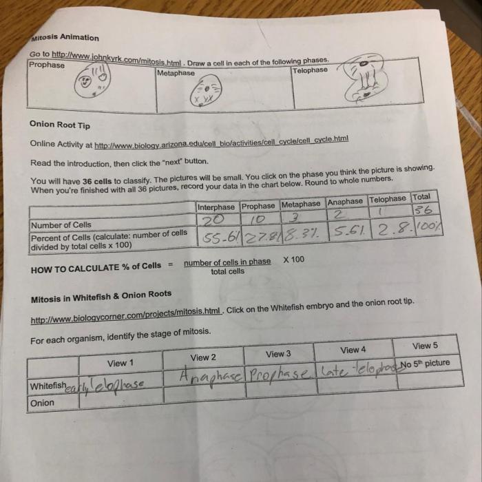
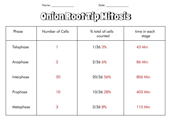
Observations and data collection during the mitosis lab are crucial for understanding the stages and characteristics of cell division. To organize and present these observations, a table or bullet points can be used.
The table should include columns for the different stages of mitosis, such as prophase, metaphase, anaphase, and telophase. Each column should record the number of cells observed in that particular stage. Additional columns can be added to capture other relevant data, such as cell shape, chromosome condensation, and spindle fiber formation.
Sample Table
| Stage of Mitosis | Cell Count | Other Observations |
|---|---|---|
| Prophase | 25 | Chromosomes condensing, nuclear membrane disappearing |
| Metaphase | 18 | Chromosomes aligned at the metaphase plate |
| Anaphase | 12 | Sister chromatids separating, moving to opposite poles |
| Telophase | 10 | Nuclear membranes reforming, chromosomes decondensing |
Discussion
The observed results of the time for mitosis lab demonstrate the dynamic process of cell division and provide valuable insights into the fundamental principles of mitosis. By comparing the observed stages to the theoretical model, we can assess the accuracy of our observations and gain a deeper understanding of the mechanisms underlying mitosis.
Significance of Observed Results
The observed results have significant implications for understanding cell biology and genetics. The accurate identification and characterization of the different stages of mitosis allow researchers to study the temporal and spatial regulation of cell division. This knowledge is essential for understanding the growth, development, and maintenance of multicellular organisms.
Comparison to Theoretical Model
The observed stages of mitosis generally align with the theoretical model, confirming the fundamental principles of cell division. The prophase, metaphase, anaphase, and telophase stages were clearly distinguishable, with the chromosomes exhibiting the expected behavior during each phase. This alignment validates the theoretical model and supports the understanding of mitosis as a highly regulated and sequential process.
Potential Sources of Error and Minimization
Several potential sources of error could affect the accuracy of the observed results. These include:
- Sampling error:Selecting a small or unrepresentative sample of cells may not accurately reflect the overall mitotic activity of the population.
- Preparation error:Improper fixation or staining techniques can distort the appearance of the chromosomes and make it difficult to identify the different stages of mitosis.
- Observer bias:Subjective interpretations of the observed cells can introduce errors in the identification and classification of mitotic stages.
To minimize these errors, it is important to use proper sampling techniques, employ standardized preparation protocols, and ensure that multiple observers independently verify the results.
Essential FAQs
What is the significance of mitosis in cell division?
Mitosis is essential for growth, tissue repair, and asexual reproduction. It ensures the equal distribution of genetic material to daughter cells, maintaining the genetic integrity of an organism.
How many stages are there in mitosis?
Mitosis consists of four distinct stages: prophase, metaphase, anaphase, and telophase. Each stage involves specific chromosome movements and structural changes.
What are the potential sources of error in a mitosis lab?
Errors can arise from improper slide preparation, inadequate staining, or misinterpretation of chromosome counts. Careful attention to methodology and experienced observation can minimize these errors.
